


Features
- One-third the cost of tabletop retinal cameras
- Proprietary image-quality evaluation helps eliminate callbacks for re-imaging
- Meets ISO 10940 optical standard
- Touchless auto-focus and image capture
- Integrated wireless capabilities help it adapt to your workflow
- High-resolution (2048 x 1536 pixels) 45-degree field of view
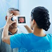
- Fast, non-mydriatic imaging to keep your patients comfortable

Easy to Use
Effective
Flexible
Secure

Seamless Workflow
Place retinal exam orders and automatically access diagnostic reports from your EMR. We offer fully integrated, bi-directional interfaces with EMRs including Allscripts®, athenahealth®, Cerner®, Epic®, NextGen® and many others to help you streamline procedures.
Watch Monica's Story
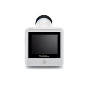
The Basics of the RetinaVue 100 Imager
Education & Documentation
Get in the know to get the most value out of your solution.
RetinaVue 100 Imager Users Guide
Product Documentation
-
Case Study
keyboard_arrow_downTelemedicine and Retinal Imaging for Improving Diabetic Retinopathy Evaluation, Case Study
-
Declaration of Conformity
keyboard_arrow_downRetinaVue Software Network; Declaration of Conformity
-
Specification Sheet
keyboard_arrow_downRetinaVue 100 Imager, Sell Sheet
-
User Manual
keyboard_arrow_downBest Practices RetinaVue® Imager and Network Client
Frequently Asked Questions
Expand All-
What is the difference between the RetinaVue 700 Imager and the 100 Imager?
keyboard_arrow_downBoth the Welch Allyn RetinaVue 100 and 700 Imagers are part of the RetinaVue care delivery model to make for simple, comfortable diabetic retinal exams in primary care. With the RetinaVue 700 Imager, this just got easier. While both imagers feature breakthrough technology with auto-focus and auto-capture, the RetinaVue 700 Imager has auto-alignment that is able to capture both eyes witout repositioning. The RetinaVue 700 Imager raises the bar to a 60 degree field of view from the 45 degree field of view of the RetinaVue 100 Imager. The RetinaVue 700 Imager also has an even smaller pupil capability at 2.5 mm (see Directions for Use) compared to the 3 mm of the RetinaVue 100 Imager.
-
Is RetinaVue Network software included with the RetinaVue 100 Imager?
keyboard_arrow_downThe RetinaVue 100 Imager is designed to work exclusively with the RetinaVue Network software. A RetinaVue Network software subscription is required and priced per camera, per month. HIPAA-compliant RetinaVue Network software enables you to easily transmit encrypted retinal images to your preferred ophthalmic specialist and manage population health data on retinal exams by clinic and patient. RetinaVue Network software subscriptions include fully integrated, bi-directional interfaces with EMRs to streamline documentation.
-
Can our preferred, local eye specialist interpret the retinal images from our clinic?
keyboard_arrow_downState-licensed, board-certified ophthalmologists and retina specialists at RetinaVue, P.C., can interpret the retinal images and prepare a comprehensive diagnostic report and referral/care plan generally in one business day—complete with ICD codes, signature and license number. RetinaVue, P.C., is the first tele-ophthalmology provider to earn The Joint Commission’s Gold Seal of Approval® by demonstrating continuous compliance with its Ambulatory Care Accreditation Standards. Professional medical services provided by RetinaVue, P.C., are priced on a per-exam basis. Alternatively, you can contract with your preferred eye specialist to interpret retinal images.
-
Does a RetinaVue Imager retinal assessment replace an annual eye exam?
keyboard_arrow_downNo. This exam only detects diseases that manifest in the retina, such as diabetic retinopathy. The RetinaVue care delivery model is not meant to be used as a replacement for a comprehensive eye exam, but rather to improve compliance for diabetic patients who are non-compliant with an annual retinal eye exam to detect retinopathy.
-
What is the RetinaVue care delivery model capable of finding?
keyboard_arrow_downWhile the exam is performed to detect and stratify the severity of diabetic retinopathy at its earliest stages, RetinaVue, P.C. retinal specialists often detect symptoms of other systemic diseases, including papilledema, macular degeneration, age-related macular degeneration, glaucoma and even certain types of cancer and stroke.
-
Are RetinaVue Imager retinal assessments reimbursed?
keyboard_arrow_downFour of the top five commercial health insurance plans reimburse for this exam when provided through the RetinaVue care delivery model. Average reimbursement is approximately $75 per exam. Check with your health plans to determine coverage and reimbursement levels, or contact one of our specialists who can assist you in this process.
-
Should I connect the RetinaVue 100 Imager via USB cable or wireless?
keyboard_arrow_downThe RetinaVue 100 Imager includes built-in wireless capabilities that enable seamless EMR connectivity and web-based control without the need for PC software. A wireless workflow is ideal if you plan to:
- Optimize your current workflow with EMR connectivity
- Use one camera across multiple clinics
- Perform exams in multiple states or in mobile environments
-
How much training is required to become proficient with the RetinaVue 100 Imager?
keyboard_arrow_downThe RetinaVue 100 Imager incorporates sophisticated technology to enhance ease of use, including touchless auto-focus, automatic image capture and proprietary image-quality evaluation. And Hillrom provides up to four hours of on-site training to ensure each clinic develops one or two personnel to expert-level proficiency.
-
Does the RetinaVue Imager retinal assessment meet the HEDIS requirement for a diabetic eye exam?
keyboard_arrow_downAbsolutely. The National Committee for Quality Assurance, which administers the HEDIS measure for diabetic eye exams, has validated that the RetinaVue care delivery model satisfies the HEDIS measure.
-
How does the image quality compare to tabletop retinal cameras?
keyboard_arrow_downLike most tabletop retinal cameras, the RetinaVue 100 Imager is capable of high-resolution (2048 x 1536 pixels) image capture, has a wide 45-degree field of view, and meets the International Organization for Standardization optical standard that specifies requirements and test methods for fundus cameras—ISO 10940.
-
How often do patients need to be dilated for a RetinaVue Imager exam?
keyboard_arrow_downApproximately 95% of patients do not require chemical dilation using the RetinaVue 100 Imager. Older patients or patients with cataracts or under certain medications may require a mild dilating drop (0.5% tropicamide).
-
Is chemical dilation required to obtain a high-quality image?
keyboard_arrow_downIn most cases, chemical dilation is not required. However, a small percentage of patients (an estimated up to 15%) may require use of a mild dilating drop (e.g., 0.5% tropicamide) to temporarily increase pupil size. There are different concentrations of dilation drops, with the mildest concentration often being enough for this exam. The effect of mild dilation drops wears off relatively quickly. Once the drops are administered, the patient should remain in a darkened room for about ten minutes before the exam.